The muscles of the back can be divided into three groups – superficial, intermediate and deep: Superficial – associated with movements of the shoulder. Intermediate – associated with movements of the thoracic cage. Deep – associated with movements of the vertebral column. The deep muscles develop embryologically in the back, and are thus described as intrinsic muscles. The superficial and intermediate muscles do not develop in the back, and are classified as extrinsic muscles. This article is about the anatomy of the superficial back muscles – their attachments, innervations and functions. The superficial back muscles are situated underneath the skin and superficial fascia. They originate from the vertebral column and attach to the bones of the shoulder – the clavicle, scapula and humerus. All these muscles are therefore associated with movements of the upper limb. The muscles in this group are the trapezius, latissimus dorsi, levator scapulae and the rhomboids. The trapezius
The muscles of the back can be divided into three groups – superficial, intermediate and deep:
- Superficial – associated with movements of the shoulder.
- Intermediate – associated with movements of the thoracic cage.
- Deep – associated with movements of the vertebral column.
The deep muscles develop embryologically in the back, and are thus described as intrinsic muscles. The superficial and intermediate muscles do not develop in the back, and are classified as extrinsic muscles.
This article is about the anatomy of the superficial back muscles – their attachments, innervations and functions.
The superficial back muscles are situated underneath the skin and superficial fascia. They originate from the vertebral column and attach to the bones of the shoulder – the clavicle, scapula and humerus. All these muscles are therefore associated with movements of the upper limb.
The muscles in this group are the trapezius, latissimus dorsi, levator scapulae and the rhomboids. The trapezius and the latissimus dorsi lie the most superficially, with the trapezius covering the rhomboids and levator scapulae.
Trapezius
it is a large, triangular-shaped muscle located in the upper back and neck region. It is named after its trapezoid-like shape and plays a crucial role in various movements of the shoulder girdle and neck. The trapezius muscle is divided into three distinct regions:
1. Upper Trapezius : This portion originates from the base of the skull and runs down to the midpoint of the spine between the shoulder blades. It helps in elevating and upwardly rotating the shoulder blades, allowing you to shrug your shoulders and tilt your head back.
2. Middle Trapezius : This part spans from the midpoint of the spine between the shoulder blades to the lower tip of the shoulder blades. It assists in retracting the shoulder blades, which involves squeezing them together. This action is commonly used when you pull your shoulders back.
3. Lower Trapezius: The lower fibers originate from the lower thoracic spine and attach to the bottom of the shoulder blade. This portion helps in depressing and upwardly rotating the shoulder blades, which occurs when you pull your shoulders down and slightly back.
- Innervation: Motor innervation is from the accessory nerve. It also receives proprioceptor fibres from C3 and C4 spinal nerves.
The trapezius muscle is involved in various movements and functions, including:
- Shrug: Elevating the shoulders.
- Scapular Retraction: Pulling the shoulder blades together.
- Scapular Depression: Pulling the shoulder blades down.
- Scapular Upward Rotation: Rotating the shoulder blades upward.
- Head Extension: Tipping the head backward.
- Neck and Upper Back Support: Stabilizing the neck and upper back during various activities.
Latissimus Dorsi
The latissimus dorsi, commonly referred to as the "lats," is a large and powerful muscle located in the back. It is responsible for a variety of movements involving the shoulder and upper arm. Here are some key points about the latissimus dorsi muscle:
1.Anatomy: The latissimus dorsi muscle is a broad, flat muscle that spans across the lower and middle back. It originates from several points, including the lower spine, the iliac crest (hip bone), and the lower ribs. It inserts into the upper arm bone (humerus) near the shoulder joint.
2. Function: The primary functions of the latissimus dorsi muscle include:
- Shoulder Extension: Pulling the upper arm downward and backward, as in activities like swimming, rowing, and pull-ups.
- Shoulder Adduction: Bringing the upper arm toward the midline of the body.
- Shoulder Medial Rotation: Internally rotating the upper arm, as in reaching across the body.
- Lumbar Extension: Assisting in arching the lower back.
- Innervation: Thoracodorsal nerve.
Levator Scapulae
The levator scapulae is a small strap-like muscle. It begins in the neck, and descends to attach to the scapula.
- Attachments: Originates from the transverse processes of the C1-C4 vertebrae and attaches to the medial border of the scapula.
- Innervation: Dorsal scapular nerve.
- Actions: Elevates the scapula.
Rhomboids
There are two rhomboid muscles – major and minor. The rhomboid minor is situated superiorly to the major.
Rhomboid Major
- Attachments: Originates from the spinous processes of T2-T5 vertebrae. Attaches to the medial border of the scapula, between the scapula spine and inferior angle.
- Innervation: Dorsal scapular nerve.
- Actions: Retracts and rotates the scapula.
Rhomboid Minor
- Attachments: Originates from the spinous processes of C7-T1 vertebrae. Attaches to the medial border of the scapula, at the level of the spine of scapula.
- Innervation: Dorsal scapular nerve.
- Actions: Retracts and rotates the scapula.
Intermediate Group
The intermediate group contains two muscles – the serratus posterior superior and serratus posterior inferior. These muscles run from the vertebral column to the ribcage, and assist with elevating and depressing the ribs. They are thought to have a slight respiratory function.
Serratus Posterior Superior
The serratus posterior superior is a thin, rectangular shaped muscle. It lies deep to the rhomboid muscles on the upper back.
- Attachments: Originates from the lower part of the ligamentum nuchae, and the cervical and thoracic spines (usually C7 – T3). The fibres pass in an inferolateral direction, attaching to ribs 2-5.
- Actions: Elevates ribs 2-5.
- Innervation: Intercostal nerves.
Serratus Posterior Inferior
The serratus posterior inferior is broad and strong. It lies underneath the latissimus dorsi.
- Attachments: Originates from the thoracic and lumbar spines (usually T11 – L3). The fibres pass in a superolateral direction, attaching to ribs 9-12.
- Actions: Depresses ribs 9-12.
- Innervation: Intercostal nerves.
Deep Muscles Group
The deep muscles develop embryologically in the back, and are thus described as intrinsic muscles. The superficial and intermediate muscles do not develop in the back, and are classified as extrinsic muscles.
This article is about the anatomy of the deep (intrinsic) back muscles – their attachments, innervations and functions.
The deep muscles of the back are well-developed, and collectively extend from the sacrum to the base of the skull. They are associated with the movements of the vertebral column, and the control of posture.
The muscles themselves are covered by deep fascia, which plays a key role in their organisation.
Anatomically, the deep back muscles can be divided into three layers; superficial, intermediate and deep. We shall now look at each layer in more detail
Superficial
The superficial muscles are also known as the spinotransversales. There are two muscles in this group – splenius capitis and splenius cervicis. They are both associated with movements of the head and neck.
They are located on the posterolateral aspect of the neck, covering the deeper neck muscles.
Splenius Capitis
- Attachments: Originates from the lower aspect of the ligamentum nuchae, and the spinous processes of C7 – T3/4 vertebrae. The fibres attach to the mastoid process and the occipital bone of the skull.
- Innervation: Posterior rami of spinal nerves C3 and C4.
- Actions: Rotate head to the same side.
Splenius Cervicis
- Attachments: Originates from the spinous processes of T3 – T6 vertebrae. The fibres attach to the transverse processes of C1-3/4.
- Innervation: Posterior rami of the lower cervical spinal nerves.
- Actions: Rotate head to the same side.
Note: The two splenius muscles can also act together to extend the head and neck.
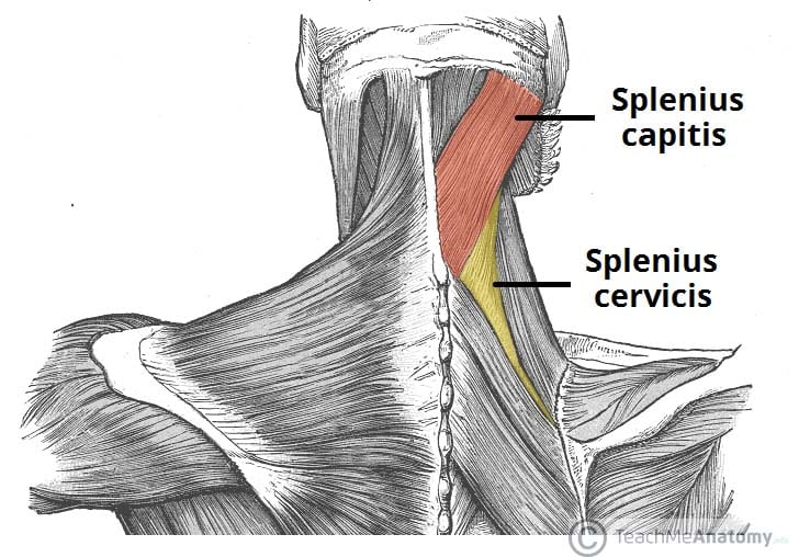
Fig 1 – The splenius muscles, located with the superficial layer of intrinsic back muscles.
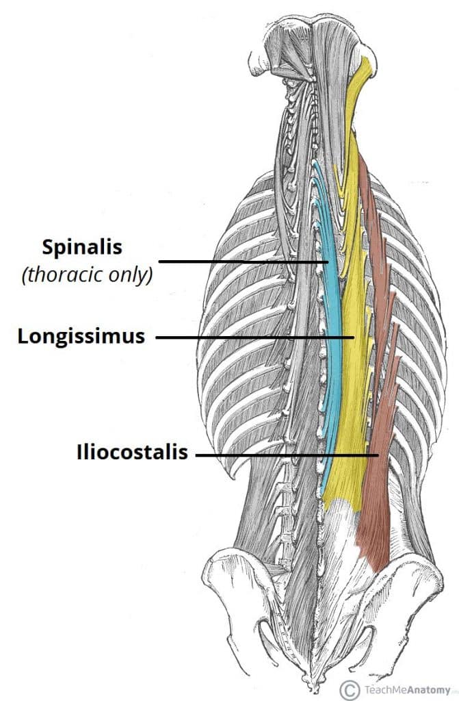
Intermediate
There are three intermediate intrinsic back muscles – the iliocostalis, longissimus and spinalis. Together these muscles form a column, known as the erector spinae.
The erector spinae is situated posterolaterally to spinal column, between the vertebral spinous processes and the costal angle of the ribs.
All three muscles can be subdivided by their superior attachments (into lumborum, thoracic, cervicis and capitis). They also all have a common tendinous origin, which arises from:
- Lower thoracic and lumbar vertebrae
- Sacrum.
- Posterior aspect of iliac crest.
- Sacroiliac and supraspinous ligaments.
Iliocostalis
The iliocostalis muscle is located laterally within the erector spinae. It is associated with the ribs, and can be divided into three parts – lumborum, thoracis, and cervicis.
- Attachments: Arises from the common tendinous origin, and attaches to the costal angle of the ribs and the cervical transverse processes.
- Innervation: Posterior rami of the spinal nerves.
- Actions: Acts unilaterally to laterally flex the vertebral column. Acts bilaterally to extend the vertebral column and head.
Longissimus
The longissimus muscle is situated between the iliocostalis and spinalis. It is the largest of the three columns. It can be divided into three parts – thoracic, cervicis and capitis.
- Attachments: Arises from the common tendinous origin, and attaches to the lower ribs, the transverse processes of C2 – T12, and the mastoid process of the skull.
- Innervation: Posterior rami of the spinal nerves.
- Actions: Acts unilaterally to laterally flex the vertebral column. Acts bilaterally to extend the vertebral column and head.
Spinalis
The spinalis muscle is located medially within the erector spinae. It is the smallest of the three muscle columns. It can be divided into the thoracic, cervicis and capitis (although the cervicis part is absent in some individuals).
- Attachments: Arises from the common tendinous origin, and attaches to the spinous processes of C2, T1-T8 and the occipital bone of the skull.
- Innervation: Posterior rami of the spinal nerves.
- Actions: Acts unilaterally to laterally flex the vertebral column. Acts bilaterally to extend the vertebral column and head.
Deep
The deep intrinsic muscles are located underneath the erector spinae, and are known collectively as the transversospinales. They are a group of short muscles, associated with the transverse and spinous processes of the vertebral column.
There are three major muscles in this group – the semispinalis, multifidus and rotatores.
Semispinalis
The semispinalis is the most superficial of the deep intrinsic muscles. Much like the intermediate muscles, it can be divided by its superior attachments into thoracic, cervicis and capitis.
- Attachments: Originates from the transverse processes of C4-T10. The fibres ascend 4-6 vertebral segments, attaching to the spinous processes of C2-T4, and to the occipital bone of the skull.
- Innervation: Posterior rami of the spinal nerves.
- Actions: Extends and contralaterally rotates the head and vertebral column.
Multifidus
The multifidus is located underneath the semispinalis muscle. It is most developed in the lumbar area.
- Attachments: Has a broad origin – arises from the sacrum, posterior iliac spine, common tendinous origin of the erector spinae, mamillary processes of lumbar vertebrae, transverse processes of T1-T3 and articular processes of C4-C7. The fibres ascend 2-4 vertebral segments, attaching to the vertebral spinous processes.
- Innervation: Posterior rami of the spinal nerves.
- Actions: Stabilises the vertebral column.
Rotatores
The rotatores are the deepest muscles of the transversospinales group. They are most prominent in the thoracic region.
- Attachments: Originates from the vertebral transverse processes. The fibres ascend and attach to the lamina and spinous processes of the vertebrae above.
- Actions: Contributes to extension and rotation of the vertebral column. Also stabilises the vertebrae and had a proprioceptive function.
- Innervation: Posterior rami of the spinal nerves
Minor Deep Intrinsic Muscles:
- Interspinales – Spans between adjacent spinous processes. Acts to stabilise the vertebral column.
- Intertranversarii – Spans between adjacent transverse processes. Acts to stabilise the vertebral column.
- Levatores costarum – Originates from the transverse processes of C7-T11, and attaches to the rib immediately below. Acts to elevate the ribs.


Comments
Post a Comment